IF WE ASKED you to list three things that happen in a typical dental exam, dental X-rays would probably be one of them. Do you know what they’re used for? It depends on the type of X-ray, so let’s look at what these are.

Getting The Wide Shot With Panoramic X-Rays
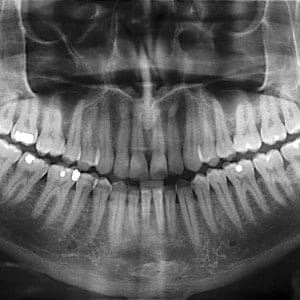
If you’ve ever stood on a circular thing with your chin on a little platform and been told to stand still for a few seconds while a machine spun around your head, you’ve had a panoramic X-ray. This is the most common type of extraoral (outside the mouth) X-ray.
Panoramic X-rays show the entire mouth in one image. In them, we can see incoming adult teeth and wisdom teeth, including impacted ones, which is how we can determine whether there is enough room for them and if they’ll come in without any extra help. This type of X-ray also makes it easier to detect abscesses, tumors, and cysts.
Enhance: Periapical X-Rays And Bitewing X-Rays
In photography, wide shots show a lot, but they aren’t as useful for details as a closeup. The same is true in X-rays, which is why we don’t rely only on the panoramic image. The next level of dental X-rays are the bitewing X-rays. These intraoral (inside the mouth) X-rays focus on a specific area inside the mouth at a time, and we usually take one for each of the four quadrants of your mouth.
Bitewing X-rays give us a better view of the gaps between teeth, which are hard to see with the naked eye. These images make it easy to check for tooth decay and cavities in those areas. When we need to get even more detailed, we take periapical X-rays, which hone in on an individual problem tooth .
Are Dental X-Rays Safe?
Dental X-rays involve brief exposure to low levels of radiation, but they are considered extremely safe. The short exposure time and protective coverings like the lead apron ensure that radiation exposure is as low as possible. We also only take X-rays as often as we absolutely need to, which further reduces exposure.
Factors that determine whether or not X-rays are necessary include the patient’s age, stage of dental development, oral health history, risk factors for various conditions, and whether or not they are presenting symptoms of oral health problems.
What is Dental Cone Beam CT?
We use Dental cone beam computed tomography (CT) , a special type of x-ray machine, in situations where regular dental x-rays are not sufficient. Our office does not use them for routine exams. This type of CT scanner uses a special type of technology to generate 3-D images of dental structures, soft tissues, nerve paths and bones in a single scan. Images obtained with cone beam CT allow for more precise treatment planning.
Patients are asked to remove all metal objects, including jewelry, eyeglasses, dentures and hairpins before having the image taken. You may also be asked to remove hearing aids and removable dentures.
With cone beam CT, an x-ray beam is moved around the patient to produce a large number of images. CT scans and cone beam CT both produce high-quality images.
A CBCT image provides detailed images of the bone and is performed to evaluate diseases of the jaw, teeth, facial bones, nasal cavity and sinuses.
We have all of these different x-ray machines available for our patients. Having multiple types of imaging systems allows for us to provide the highest quality dental care available for our patients.
Still Have X-Ray Questions?
If you would like to know more about how we use dental X-rays in our practice or have any concerns about safety, just ask us! We want our patients to have all the information they need to feel comfortable when they come to see us.
Your dental health is in good hands with All About You Dental Care!


